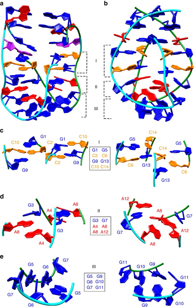
Figure 8. Comparison of VK34_I11 and VK1 structures.
(a) Overall representation of VK34_I11 structure. (b) VK1 structure. (c) Side views of G-C base pairs in VK34_I11 (left) and VK1 (right). (d) Insight into two G-A base pairs and the two A4 (left) and two A8 (right) residues orientated inside a hydrophobic pocket in VK34_I11 (left) and VK1 (right). (e) Side view of three G-G base pairs in fold-back arrangements in VK34_I11 (left) and VK1 (right). The roman numerals I, II and III refer to locations of the individual regions within the overall structure shown in a. The guanine residues are coloured blue, adenine red, cytosine orange and inosine purple. The two strands are coloured green and cyan.

