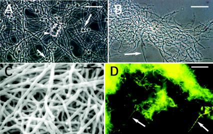
FIG. 1.
Morphology of some of the actinobacteria analyzed in this report. (A) Casuarina strain CcI3 with multilocular sporangia (arrowhead) and diazovesicles (arrows) grown in liquid BAP minus N medium. Bar, 20 μm. (B) Hyphae (arrow) of L5 from a culture grown in stirred BAP plus N. Bar, 40 μm. (C) Scanning electron micrograph of hyphae from strain 8103 grown in stirred BAP plus N. Bar, 2.5 μm. (D) Hyphae of 7702 stained with acridine orange. Segmented hyphae are indicated with the arrow and in the insert. Bar, 20 μm.

