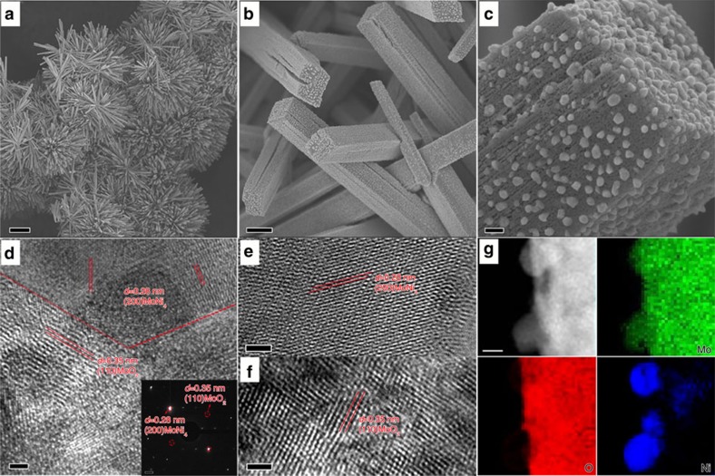
Figure 2. Morphology and chemical composition analyses of MoNi4/MoO2@Ni.
(a–c) Typical SEM and (d–f) HRTEM images of MoNi4/MoO2@Ni; (g) corresponding elemental mapping images of the MoNi4 electrocatalyst and the MoO2 cuboids. The inset image in d is the related selected-area electron diffraction pattern of the MoNi4 electrocatalyst and the MoO2 cuboids. Scale bars, (a) 20 μm; (b) 1 μm; (c) 100 nm; (d–f) 2 nm; inset in d, 1 1/nm; (g) 20 nm.

