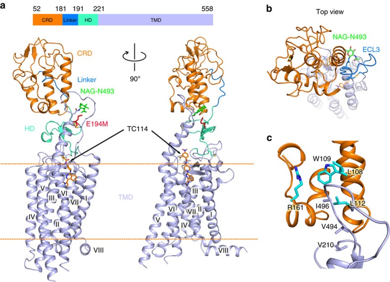
Figure 2. Overall structure of the multi-domain human SMO.
(a) Overall structure of the human SMO in complex with TC114 determined at an XFEL. TC114 is shown as orange sticks. The CRD, linker, HD and TMD are indicated as orange, marine, green cyan and light blue cartoons, respectively. The membrane boundary is labelled, as an orange dashed line. N-linked glycans (NAG) are shown in green sticks. (b) Top view of the SMO from the extracellular side. A hydrophobic pocket is formed by the CRD hydrophobic groove and ECL3 (marine loop). (c) Key residues in the CRD and ECL3 defining the hydrophobic pocket are shown in cyan and light blue sticks, respectively.

