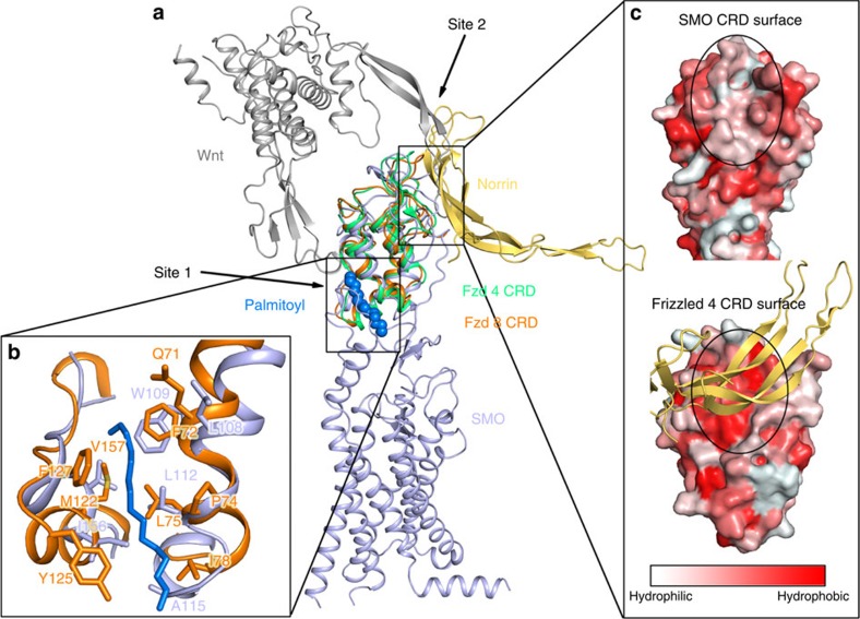
Figure 4. Comparison of the CRD of the human SMO with the Frizzled receptors.
(a) Side view of superimposed structures of the human SMO (hSMO) CRD with the hFzd-4 (PDB ID: 5CL1) and mFzd-8 CRDs (PDB ID: 4F0A). The SMO, hFzd-4 and mFzd-8 are shown as cartoons in light blue, lime green and orange, respectively. The Wnt and Norrin are shown as dark grey and yellow orange cartoons, respectively. The palmitoyl group in the mFzd-8 CRD is shown in marine spheres. (b) Site 1: Close-up view of the palmitoyl group with interacting residues as orange sticks. The palmitoyl group is shown in marine sticks. The residues from SMO forming the hydrophobic pocket are shown in light blue sticks, mFzd-8 in orange sticks. (c) Site 2: Surface of SMO and hFzd-4 CRD. Norrin is shown in light orange cartoons. The colour gradient from light red to dark red corresponds to the change of surface property from hydrophilic to hydrophobic. The Norrin binding site on hFzd-4 CRD surface is labelled by a black dashed circle with corresponding site also marked on the SMO surface.

