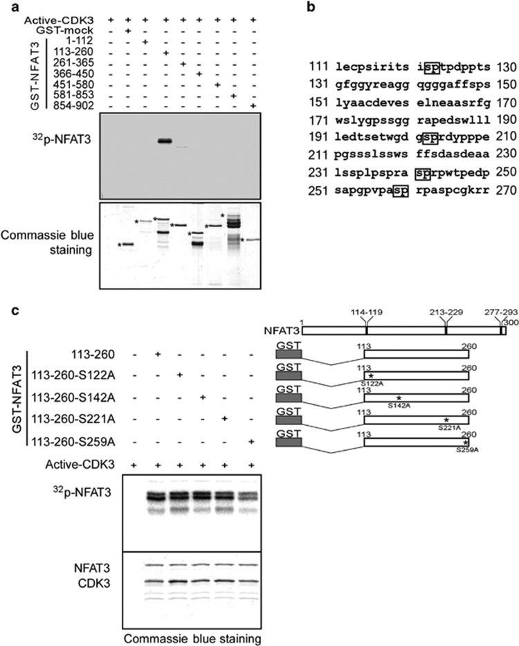
Figure 4.
CDK3 phosphorylates NFAT3 at Ser259. (a) GST-tagged truncated NFAT3 was used for an in vitro kinase assay with active CDK3. The phosphorylation was visualized by autoradiography (32P-NFAT3). Coomassie blue staining was used to verify equal loading of GST-tagged truncated NFAT3 proteins (labeled with *). (b) Sequence of NFAT3 from amino acids 111–260. The predicted SP motifs are indicated with black boxes. (c) GST-tagged wildtype NFAT3 fragments or GST-tagged mutant NFAT3 fragments were used for an in vitro kinase assay with active CDK3 as indicated (upper panel). The phosphorylation was visualized by autoradiography (32P-NFAT3) and Coomassie blue staining indicated the CDK3 protein and respective NFAT3 fragments (indicated as NFAT3 and CDK3).

