Figure 7.
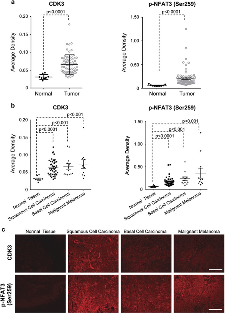
CDK3 and phosphorylated NFAT3 are overexpressed in human skin cancer. (a) Immunofluorescence staining was used to investigate the expression of CDK3 and phosphorylated NFAT3 (Ser259) in human skin cancer (n=65) and normal tissues (n=9). The staining was evaluated by average density and used for statistical analysis (P<0.0001). (b) Tumor tissues were further divided by subtype: squamous cell carcinoma (n=41), basal cell carcinoma (n=13) and malignant melanoma (n=11). The average density was calculated by dividing the integrated density by the positive area. The data was presented as mean values of intensity score and P-value (Mann–Whitney U test). (c) Representative photos of immunofluorescence staining performed on the SK801b skin cancer tissue arrays as described in ‘Materials and methods' section. Tissues were incubated with antibodies to detect CDK3 or phosphorylated NFAT3 (Ser259). A secondary antibody conjugated with Alexa Fluor 647 (red for CDK3 or phosphorylated NFAT3) was used for final detection. Scale bar, 100 μm.

