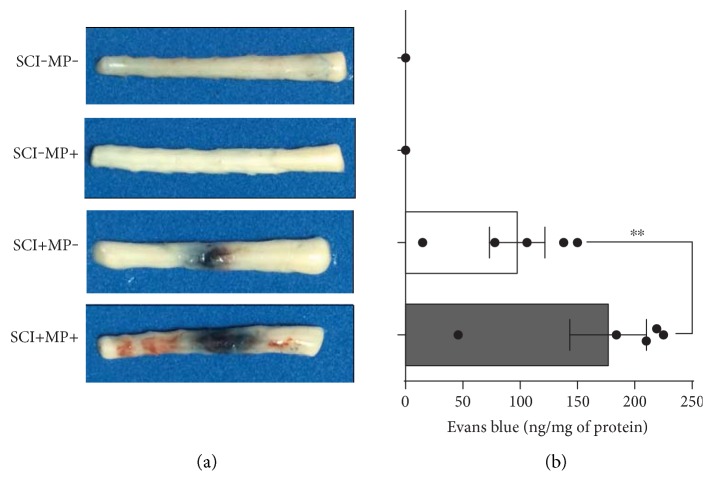
Figure 2.
Evans blue extravasation is increased in rats subjected to SCI and treated with MP. (a) Representative photographs of spinal cord segments T5-6 to L1-2 showing the lesion caused by SCI 24 h after contusion and the extravasated Evans blue (n = 5 per group). Sham-operated animals do not have any trace of Evans blue in the spinal cord parenchyma; however, the accumulation of this tracer at the site of injury is evident in animals subjected to SCI. MP worsen BSCB disruption causing a further accumulation of the dye at the site of injury. (b) Fluorescence quantification of the extravasated Evans blue shows a notable increase of ~80% parenchymal Evans blue in animals treated with MP related to injured rats administered with vehicle alone. Bars are the mean ± SEM of 5 spinal cord segments; dots show individual data measurements. ∗∗p < 0.01.

