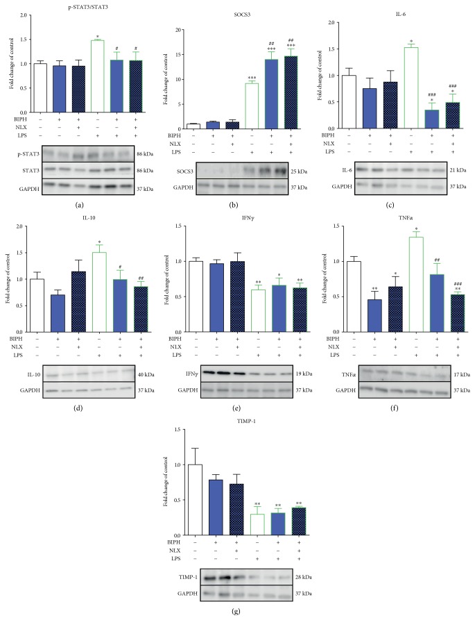
Figure 5.
The influence of biphalin on STAT3 (a) phosphorylation and SOCS3 (b), IL-6 (c), IL-10 (d), IFNγ (e), TNFα (f), and TIMP-1 (g) protein levels in vehicle- and LPS-treated primary microglial cells. Microglial cells were treated with biphalin (BIPH; 10 μM) for 30 min and then with LPS (100 ng/mL) for 1 h (a) or 24 h (b, c, d, e, f, g). Naloxone (NLX; 0.1 μM) was added 30 min before biphalin. The data are presented as the fold change compared with the control group (vehicle-treated cells) as the mean ± SEM of 3-4 independent experiments. The results were statistically evaluated using one-way analysis of variance (ANOVA) followed by Bonferroni's post hoc test to assess differences between the treatment groups. Significant differences in comparison with those of the control group (vehicle-treated cells) are indicated by ∗P < 0.05, ∗∗P < 0.01, and ∗∗∗P < 0.001; differences between LPS-treated and biphalin- or biphalin- and naloxone-treated cells are indicated by #P < 0.05, ##P < 0.01, and ###P < 0.001.

