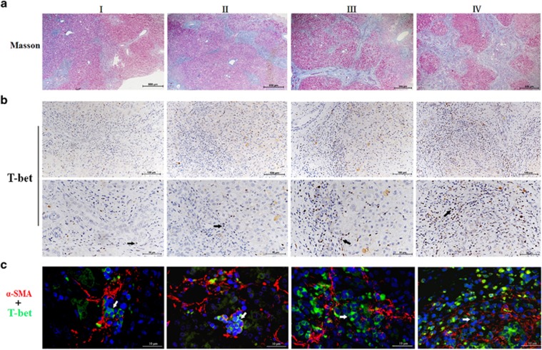
Figure 3.
Intrahepatic Th1 cells were linked to the progress of liver fibrosis in BA. (a) Successive liver sections were used for Masson's trichrome staining to assess the stage of liver fibrosis. Data shown are representative of 32 early-stage BA patients with liver fibrosis grades I to IV. (b) Immumohistochemical staining was performed to detect T-bet+ cells, which specifically identifies Th1 cells.Black arrows indicate T-bet+Th1 cells. (c) Immunofluorescence staining for alpha-SMA+HSCs (red) and T-bet+Th1 cells (green) identified HSCs surrounded by Th1 cells. White arrows mark the positions of T-bet+Th1 cells

