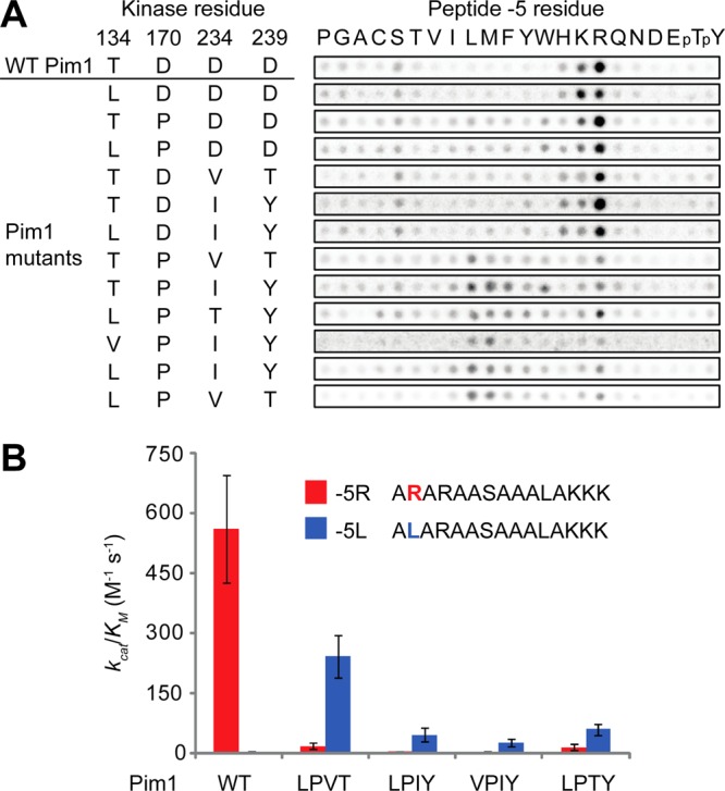
Figure 2.

Re-engineering of the phosphorylation site specificity of Pim1. (A) Peptide array analysis of Pim1 mutants showing specificity at the −5 position. Spot intensities reflect the extent of phosphorylation using radiolabeled ATP. pT, phosphothreonine; pY, phosphotyrosine. (B) Quantitative phosphorylation parameters for Pim1 and quadruple mutants on peptide substrates assessed by radiolabel kinase assay. LPVT, Pim1-T134L/D170P/D234 V/D239T; LPIY, Pim1-T134L/D170P/D234I/D239Y; VPIY, Pim1-T134 V/D170P/D234I/D239Y; LPTY, Pim1-T134L/D170P/D234T/D239Y. Error bars indicate standard deviation (n = 3).
