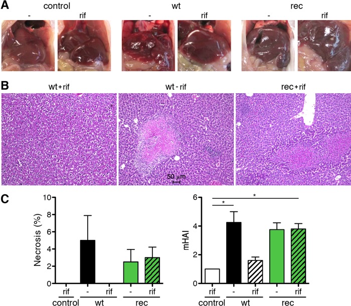
FIG 6.
Comparable liver pathology in R. typhiGFPuv- and wild-type R. typhi-infected CB17 SCID mice. (A) Representative images of the livers of the animals shown in Fig. 4 and harvested from moribund mice after euthanasia demonstrate liver necrosis in mice infected with wild-type R. typhi in the absence of antibiotics and in animals that were infected with R. typhiGFPuv, whether treated with rifampin (rif) or not (−). Livers of control mice that received PBS or were infected with wild-type R. typhi and were treated with rifampin appeared healthy. (B) Representative H&E staining of the liver of a rifampin-treated mouse infected with wild-type R. typhi (left), a mouse infected with wild-type R. typhi without antibiotic treatment (center), and a rifampin-treated mouse infected with R. typhiGFPuv (right). Livers from rifampin-treated animals infected with wild-type R. typhi appeared healthy, while necrotic lesions and cellular infiltrates were detectable in the livers from untreated mice infected with wild-type R. typhi, as well as in mice infected with R. typhiGFPuv. (C) The percentages of necrotic areas (left) and the mHAI were quantified. The graphs show means and SEM. Statistical analysis was performed using one-way ANOVA (Kruskall-Wallis test followed by Dunn's posttest). The asterisks indicate statistically significant differences (*, P < 0.05).

