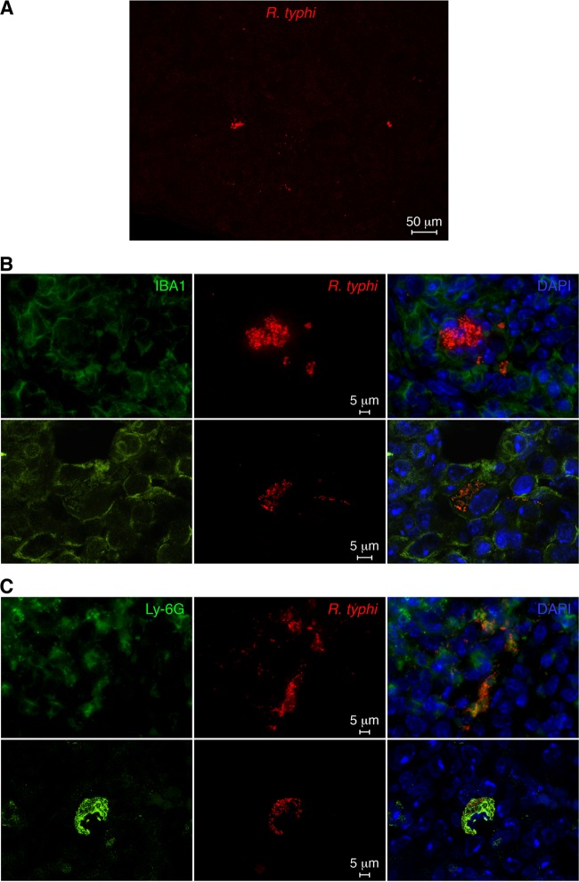
FIG 7.
Detection of R. typhiGFPuv in histological sections of the spleen from a moribund infected CB17 SCID mouse. (A) R. typhiGFPuv bacteria were detected in spleen sections from a moribund infected CB17 SCID mouse by staining with anti-GFP antibody (red). An overview image is shown. (B) Sections were further stained with DAPI (nucleus; blue) and for IBA1 (green). Shown are confocal images (bottom) and overlays (right). (C) In addition, sections were stained with DAPI (blue) and for Ly-6G (green). Shown are confocal images (bottom) and overlays (right).

