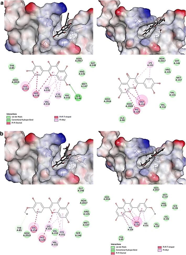
Fig. 5.

Molecular docking and interaction images of compounds 2 and 4 with Keap1. a Quercetin docked in BTB domain of Keap1 (left-up side) and mutant BTB domain at C151W of Keap1 (right-up side). The interpolated structures were shown in this figure. The 2D diagram of ligand interactions was illustrated at down side. b Quercetin-4′-methyl ether docked in BTB domain of Keap1 (left-up side) and mutant BTB domain at C151W of Keap1 (right-up side). The interpolated structures were shown in this figure. The 2D diagram of ligand interactions was illustrated at down side
