Figure 1.

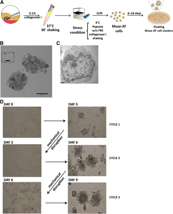
Generation and culture of liposuction‐derived Muse‐AT cells. (A): Scheme of Muse‐AT cell preparation protocol. Muse‐AT cells were obtained after digestion with collagenase and severe stress conditions. Clusters were generated when Muse‐AT cells were seeded on nonadherent plastic. (B): Muse‐AT cells formed clusters of 50–150 µm in diameter that grew in suspension culture. Scale bar = 50 µm. Left inset, black arrow indicates a single Muse‐AT cell at the edge of a cluster. Scale bar = 20 µm. (C): Transmission electron microscopy of a representative Muse‐AT cell after 5–7 days in culture showing a high nucleus to cytoplasm ratio. Scale bar = 500 nm. (D): Muse‐AT cells were seeded immediately on nonadhesive dishes after isolation (day 0) and formed clusters after 3 days of culture (day 3, cycle 1). After mechanical disaggregation, spheroids formed again, reaching 50–150 µm in diameter during the second and third growth cycle. Abbreviations: FBS, fetal bovine serum; Muse‐AT, multilineage‐differentiating stress‐enduring cells derived from adipose tissue; O/N, overnight; w/o, without.
