Figure 4.

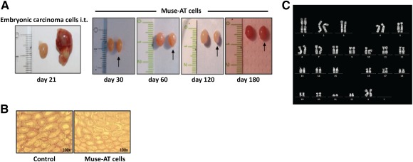
Lack of teratoma formation after transplantation and stability of Muse‐AT cells. (A): Muse‐AT cells were injected (106) i.t. into NODscid mice. The transplanted mice were monitored weekly for the appearance of tumors. The P19 embryonic carcinoma cell line was injected (106) as the control. The mice were sacrificed when the tumors became outwardly apparent. NODscid mice injected with Muse‐AT cells did not develop teratoma during the observed period (up to 6 months). (B): H&E staining of testes showed normal tissue structure in NODscid Muse‐AT cells of the injected mice. (C): Representative normal karyotype of Muse‐AT cells that showed expression of stem cell markers. Abbreviations: d, day; i.t., intratesticular; Muse‐AT, multilineage‐differentiating stress‐enduring cells derived from adipose tissue.
