
Figure 2.
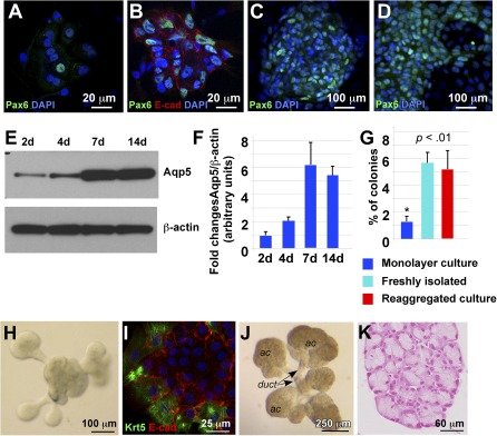
Differentiation of lacrimal gland (LG) epithelial cells in monolayer culture. (A–D): One day (A), 1 week (B, C), and 3 weeks (D). (A–D): Cultured cells were stained with antibody to Pax6 (green). (B): Cells were stained with antibodies to Pax6 (green) and E‐Cadherin (red). Nuclei are stained with DAPI (blue). (E): Western blot showing expression of aquaporin‐5 (Aqp5) in fluorescence‐activated cell sorting‐isolated epithelial progenitor cells (EPCPs) cultured for 2, 4, 7, and 14 days. (F): Quantification of protein abundance as measured from immunoblot band intensities (e.g., E) in three independent experiments. Values represent the optical density of Aqp5 bands normalized to that of β‐actin bands (means of three experiments ± SD). (G): Comparison of colony forming abilities (using an in vitro colony forming efficiency [CFE] assay) of EPCPs grown in monolayer, freshly isolated EPCPs, and EPCPs grown in three‐dimensional (3D) reaggregated cultures. The CFE was calculated as the number of colonies at day 8 as a proportion of the number of cells plated in a well. In each experiment, six replicate wells were analyzed per each experimental condition. The final CFE was calculated in three independent experiments (i.e., a total of 18 wells per each condition). p value was determined versus control (freshly isolated EPCPs). Asterisk indicates significant differences between monolayer cultures and freshly isolated or reaggregated cultures: *, p < .01. (H): EPCPs grown in 3D reaggregated culture form buds in 24–48 hours. (I): Buds express Krt5 (green) and E‐cadherin (red). (J): LG reaggregates cultured for 1 week differentiate into ductal and acinar components. (K): Transverse section of one of the acinar components of the 3D reaggregated culture showing acinar structure. Section was stained with Fast red to visualize nuclei. Abbreviations: ac, acinar; DAPI, 4′,6‐diamidino‐2‐phenylindole; duct, ductal; E‐cad, E‐cadherin.