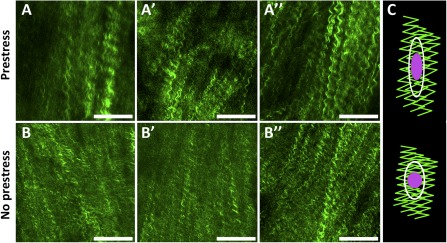
Figure 4.

Representative images of collagen in periosteal fibrous layer immediately after harvest with prestress (A, A′, A″) and without prestress (B, B′, B″), and schematic of working hypothesis (C). The working hypothesis was that loss of intrinsic tissue prestress and concomitant relaxation in collagen crimping at fibrous layer would lead to cell nucleus shape change (rounding) at the cambium layer. Collagen is represented in green, cell nucleus in purple, and cell boundary in white. Scale bars = 100 μm.
