
Figure 2.
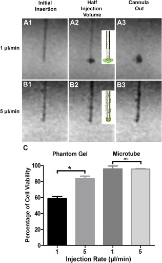
Impact of infusion rate and physical constraints on cell dispersion and survival in a brain phantom gel. Magnetic resonance imaging scans showing the progression of superparamagnetic iron oxide‐labeled neural stem cells (NSCs) infused into agarose gel phantoms at 1 μl per minute (A1–A3) and 5 μl per minute (B1–B3). The schematic drawings (A2, B2) depict the flow and dispersion (red arrows) of cells (1 μl per minute) (A2) and backflow of cells up the cannula trajectory (5 μl per minute) (B2). The postinjection scans (cannula out) show a well‐formed cloud of NSCs at 1 μl per minute (A3) and backflow of NSCs along the cannula trajectory at 5 μl per minute (B3). (C): NSCs injected either into the phantom gel or into the air inside a microtube show that slow injection (1 µl per minute) causes more cell death than does faster injection rate (5 µl per minute), although no significant change in viability was observed when NSCs were injected into a microtube. Data represent the mean ± SEM of experiments performed in triplicate in three independent experiments. ∗, p < .05. Abbreviation: ns, not significant.