Figure 4.

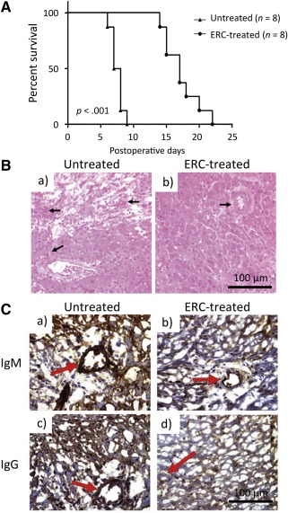
ERCs attenuate murine cardiac allograft rejection. (A): Prolonged murine cardiac allograft survival of ERC‐treated recipients. C57BL/6 (H‐2b) hearts were heterotopically transplanted to BALB/c (H‐2d) mice. In treated groups, recipients were injected i.v. with ERCs (1 × 10⁶ cells per mouse) 24 hours after cardiac transplantation. Beating of the grafted heart was monitored daily by direct abdominal palpation. End‐point for each animal represents cessation of cardiac impulses (n = 8). p value was determined by log‐rank survival test (∗, p < .001, survival proportions of recipients treated with ERCs vs. survival proportions of recipients without treatment). (B): Histology of cardiac allografts recipients. On postoperative day 8, the rejection time‐point for the untreated group, grafts were harvested and evaluated by H&E staining of paraffin sections (n = 8). (Ba): Cardiac grafts from untreated transplant recipients. (Bb): Grafts from ERC‐treated recipients. Arrows indicate intravascular and/or interstitial changes in heart grafts. (C): Intragraft IgM and IgG deposition in cardiac transplant recipients. Antibody deposition in grafted hearts at the time of sacrifice was observed by immunohistochemistry specific for either IgM or IgG (n = 8). (Ca): Cardiac grafts from untreated transplant recipients. (Cb): Grafts from ERC‐treated recipients. Arrows indicate IgM deposition around vessels. (Cc): Grafts from untreated transplant recipients. (Cd): Grafts from ERC‐treated recipients. Arrows indicate IgG deposition around vessels. Scale bars = 100 μm. Abbreviation: ERC, endometrial regenerative cell.
