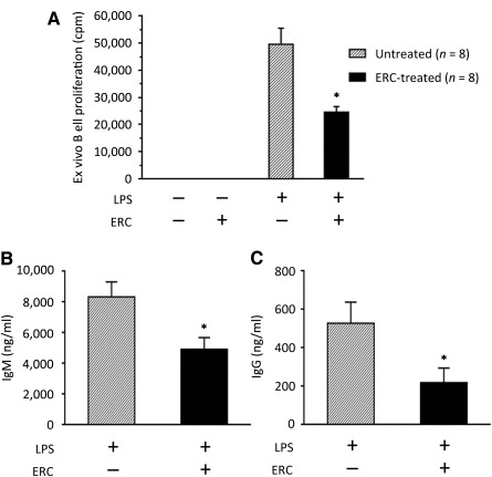
Figure 5.

Ex vivo proliferation and IgM and IgG production of ERC‐treated recipient B cells after cardiac allografting. C57BL/6 (H‐2b) hearts were heterotopically transplanted to BALB/c (H‐2d) mice. In treated groups, recipients were injected i.v. with ERCs (1 × 10⁶ cells per mouse) 24 hours after cardiac transplantation. On postoperative day 8, the rejection time point for the untreated group, mice were sacrificed and recipient splenocytes were harvested (n = 8). (A): Purified recipient B cells (105 per well) were stimulated in culture with 2 μg/ml LPS. After 48 hours of culture, 1 μCi of 3H‐thymidine was added. Eighteen hours thereafter, cells were harvested and 3H‐thymidine incorporation was measured. ∗, p < .001 vs. the proliferation of B cells from LPS‐stimulated, untreated recipients. (B, C): Purified recipient B cells (5 × 10⁵ per well) were stimulated in culture with 2 μg/ml LPS. After 6 days of culture, supernatants were harvested and (B) IgM or (C) IgG production was measured by enzyme‐linked immunosorbent assay. ∗, p < .001 vs. the levels of (B) IgM or (C) IgG antibody in the supernatants of B cells from untreated recipients. (A–C): Graphs represent mean ± SE of triplicate wells. p value was determined by a two‐tailed, paired t test. Data shown represent three sets of transplants and subsequently three separate ex vivo experiments. Abbreviations: cpm, count(s) per minute; ERC, endometrial regenerative cell; LPS, lipopolysaccharide.
