
Figure 3.
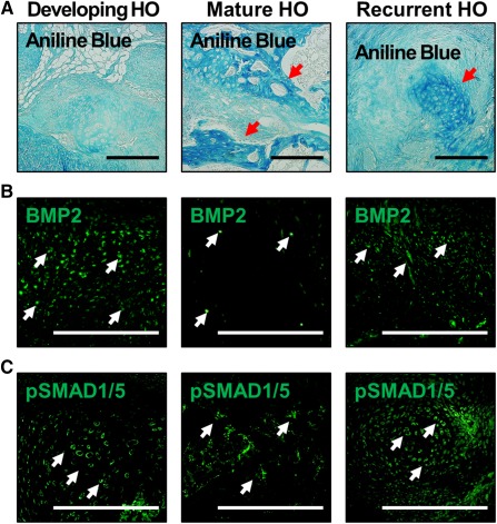
Osteoid and BMP signaling characteristics of recurrent HO. (A): Aniline blue staining shows presence of mature osteoid 9 weeks after HO, and emerging aniline blue staining in both early HO and developing recurrent HO lesions. Red arrows = sites of osteoid. (B): BMP2 expression is elevated in the developing HO lesion, reduced in mature osteoid 9 weeks after injury, and recurs in the recurrent HO lesion. (C): pSMAD 1/5 expression is elevated in the developing HO lesion, reduced in mature osteoid 9 weeks after injury, and recurs in the recurrent HO lesion. White arrows = representative sites of positive staining. Scale bar = 200 µm. Abbreviations: BMP2, bone morphogenetic protein‐2; HO, heterotopic ossification.