Figure 3.

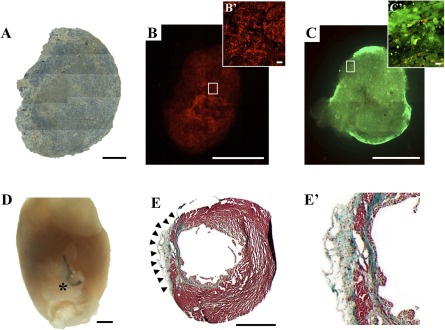
Macroscopic evaluation of the three‐dimensional engineered construct with cardiac adipose tissue‐derived progenitor cells (cardiac ATDPCs) cultured for 21 days before in vivo grafting. (A): Brightfield image composition of the cellular fibrin patch under standard culture conditions. (B, B′): PKH26 cell labeling (red) of cardiac ATDPCs in the fibrin patch (B) and magnification of the core region (B′). (C): Representative image showing cell viability (green cells, alive; red cells, dead) in a fibrin patch loaded with cardiac ATDPCs, as performed by the live/dead assay. (C′): Magnification of a construct border zone with an abundance of viable cells (green). (D): Representative photograph of an excised heart from a postinfarction animal (visible ligation) 21 days after the cellular fibrin construct implantation (asterisk). (E, E′): Representative image of Masson’s trichromic staining of heart cross‐section from a myocardial infarction plus electromechanically conditioned‐treated animal (E) and its magnification (E′). Scale bars = 1 mm (A–E) and 20 µm (B′, C′).
