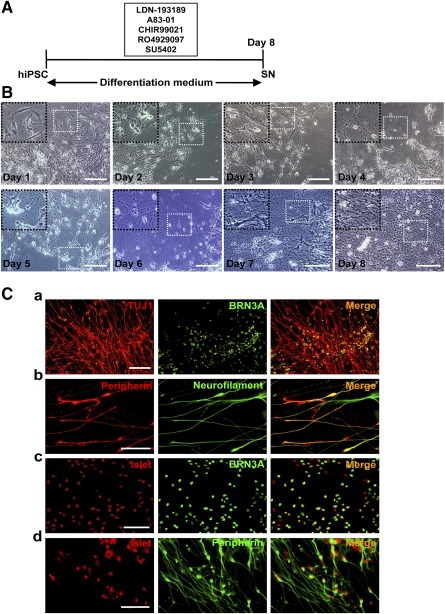
Figure 1.

One‐step protocol for the differentiation of hiPSCs to sensory neurons. (A): Schematic outline of the protocol. (B): Morphological changes in the progression from hiPSCs to sensory neurons as viewed under phase‐contrast microscopy. Scale bars = 50 μm. (C): Neuronal lineage marker expression by day‐8 cells as detected by double immunofluorescence for TUJ1 and BRN3A (Ca), peripherin and neurofilament (Cb), Islet1 and BRN3A (Cc), and Islet1 and peripherin (Cd). Scale bars = 50 μm. Abbreviations: hiPSC, human induced pluripotent stem cell; SN, sensory neuron.
