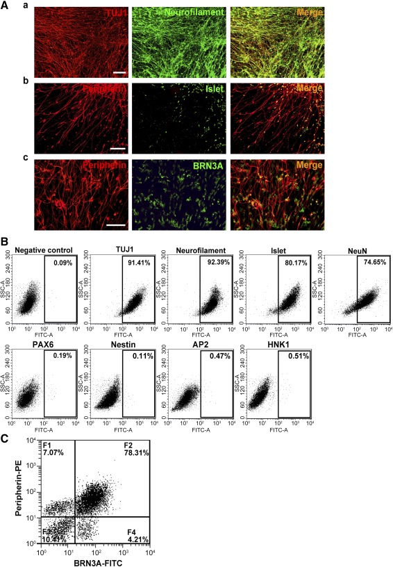
Figure 2.

Continuing culture of human iPSC‐derived neurons in maintenance medium for 14 days. (A): Neuronal lineage marker expression by the iPSC‐derived neurons as detected by double immunofluorescence for TUJ1 and neurofilament (Aa), peripherin and Islet1 (Ab), and peripherin and BRN3A (Ac). Scale bars = 50 μm. (B): Representative dot plots of flow cytometric data showing the percentage of iPSC‐derived cells positive for TUJ1 (91.41%), neurofilament (92.39%), Islet1 (80.17%), and NeuN (74.65%) but not for PAX6, Nestin, AP2, and HNK1 (<1%). (C): Representative dot plot of two‐color flow cytometric data showing subpopulations of the iPSC‐derived cells: F1, low in BRN3A but high in peripherin; F2, high in both BRN3A and peripherin; F3, low in both BRN3A and peripherin; F4, high in BRN3A but low in peripherin. Abbreviations: FITC, fluorescein isothiocyanate; iPSC, induced pluripotent stem cell; PE, phycoerythrin; SSC, side scatter.
