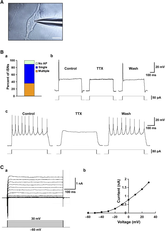
Figure 5.

Electrophysiological properties of the human iPSC‐derived sensory neurons. (A): Photomicrograph showing a patch pipette attached to an iPSC‐derived neuron. Scale bar = 10 μm. (B): Current‐clamp recording of membrane potential in iPSC‐derived neurons: proportion of neurons showing null/single/multiple action potential firing as evoked with step‐current injection (Ba); example recording of a single action potential evoked by current injection of 50 pA for 1 second (Bb); and example recording of multiple action potential firing as evoked by current injection of 80 pA for 1 second (Bc). Application of TTX blocked the generation of action potentials, but after washout, action potential firing could again be evoked. (C): Voltage‐clamp recording of specific ion currents: example recording of delayed rectifier K+ current evoked by depolarizing steps ranging from −60 mV to +30 mV (Ca); and steady‐state current‐voltage relationship for the cell examined (Cb). Abbreviations: AP, action potential; iPSC, induced pluripotent stem cell; iSN, induced sensory neuron; TTX, tetrodotoxin.
