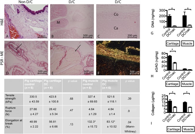
Figure 2.

Evaluation of the decellularized porcine larynx using histological, molecular, and biomechanical analyses. Sections were stained with H&E and compared against control tissue (A) for the absence of intact nuclear material within muscle (B), cartilage, and overlying collagen (C). Sections were further stained with PSE‐ME to illustrate intact elastin within the blood vessels (arrow) (D) and intact collagen fibers between the muscle bundles (arrow) (E). (F): Under polarized light, the structural integrity of the collagen was further verified. Molecular analysis for the amount of DNA, glycosaminoglycans (GAGs), and total collagen remaining within the decellularized larynx was undertaken for cartilage and muscle independently (mean ± SD; p value determined by 1‐way analysis of variance with multiple comparisons; n = 6 for each group). Statistical analysis was performed using Student's t test, for which p < .05 was considered statistically significant. The molecular data for each were as follows: cartilage: ∗, p = .0001; muscle: ∗, p = .0329 (G); cartilage: ∗, p = .0001; muscle: ∗, p = .1099 (not significant) (H); and cartilage: ∗, p = .002; muscle: ∗, p = .0001 (I). (G–I): Both decellularized cartilage (p < .05) and muscle (p < .05) showed a statistically significant reduction in the amount of DNA. Similarly, the amount of GAGs remaining with the decellularized cartilage (p < .05) was reduced but preserved within decellularized muscle tissue. Collagen content was also significantly reduced in the decellularized cartilage and muscle tissue (p < .05 for each). Biomechanical analysis of the decellularized tissue components only revealed changes for the muscle but not cartilage. Abbreviations: CA, cartilage; CO, collagen; DC, decellularized; DC vac, decellularized using vacuum technology; H&E, hematoxylin and eosin stain; M, muscle; PSR‐ME, Picrosirius red with Miller's elastin; sGAG, sulfated glycosaminoglycan.
