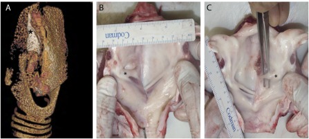
Figure 4.

Ex vivo assessment of the implanted scaffold. (A): Static computed tomography image showing the remnants of the cartilage component (marked with an asterisk) of the decellularized scaffold; a three‐dimensional rotating image is presented in supplemental online Video 1. Representative images of the internal luminal surface showing good mucosal coverage and the development of a vocal fold (marked with an asterisk) (B) and a vocal “strap” of tissue (C), both craniocaudally offset from the normal contralateral true vocal fold.
