Figure 5.

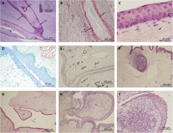
Epithelialization and mucosal coverage. (A): Remains of the tissue‐engineered oral mucosal sheet from animal 417 showing a concentration of inflammatory cells (insert) surrounding a mineralized center. (B): The collagen fibers of the original decellularized dermal collagen sheet (arrow heads) are more easily identifiable on the Picrosirius red with Miller's elastin‐stained section. In this image, the sheet has folded on itself and the paler central portion represents the original seeded aspect. Epithelialization of the mucosal surface showing stratified columnar epithelium (C) of porcine origin (insert shows positive stained human small intestine) (D). (E): The underlying mucosa shows well established vasculature (α‐SMA stain). Both vocal folds (G) and strap (H) were identified histologically. (I): Tips of the vocal folds. Abbreviations: M, mineralized; vf, vocal fold; vs, vocal strap.
