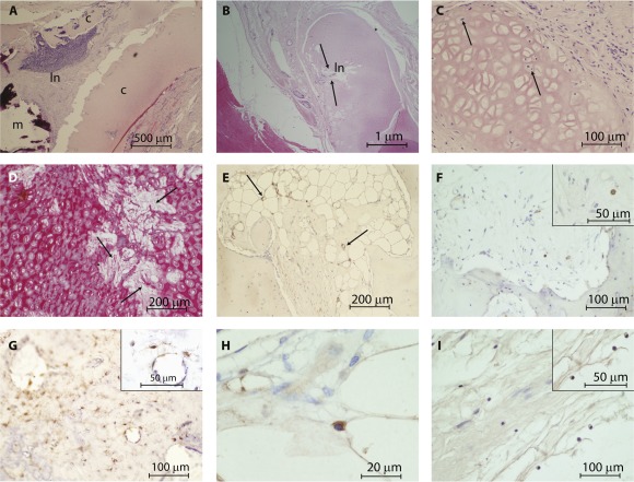
Figure 6.

Cartilage remodeling. (A): Decellularized cartilage showing infiltration by a lymph node and mineralization, resulting in degradation. (B, C): This profile was not consistent across all animals; in some cases, the acellular cartilage was infiltrated with cells (arrows). (D): In addition, where cells had infiltrated the acellular cartilage, the cartilage was slowly remodeled into collagen fibrils (arrow). (E): Where cartilage was being remodeled, as in animal 412, the original decellularized cartilage had established blood vessels (stained with α‐SMA; arrows) and cells positive for collagen II (F; insert shows cytoplasmic positivity of a cell at higher magnification) CD 44 (G; insert shows a cell at higher magnification), and collagen X (H; cytoplasmic positive cell within the extracellular matrix). (I): Potential load‐bearing changes in the cartilage were illustrated by the presence of aggrecan cytoplasmic positive cells. Abbreviations: c, cartilage; ln, lymph node; m, mineralization.
