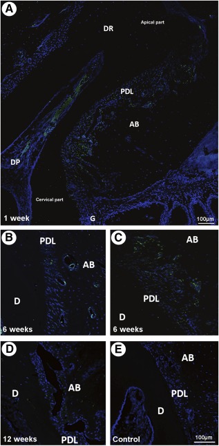
Figure 1.

Localization of the grafted green fluorescent protein (GFP)+/adipose stromal cells (ASCs). Cells were tracked by immunofluorescence. (A): GFP+/ASC were identified in the experimental, periodontium‐implanted site at 1 week, not only close to the wound bed near the cervical part but also toward the apical part of the periodontal ligament (PDL) and surrounding the ligament and alveolar bone blood vessels. (B, C): GFP+/ASC localization surrounding PDL and alveolar bone blood vessels (B) and in the apical part of the PDL (C). (D): Undistinguishable GFP+/ASC in the grafted side at week 12. (E): Undistinguishable cells in vehicle‐only treated control sites. Scale bar = 100 µm. Abbreviations: AB, alveolar bone; D, dentin; DP, dental pulp; DR, dental root; G, gingiva; PDL, periodontal ligament.
