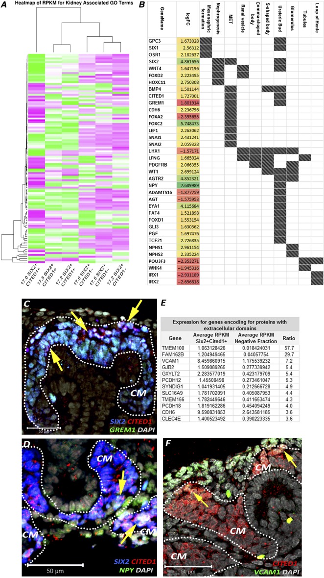
Figure 4.

Differential gene expression in SIX2+CITED1+ cells from human fetal kidney (hFK). (A): Heat map showing relative expression (measured in RPKM) in SIX2+CITED1+ cells from hFK and negative fraction for genes involved in nephrogenesis, induction, specification, and differentiation. (B): Differentially expressed genes (logFC, p < .05) involved in nephron development in SIX2+CITED1+ cells (specific renal developmental processes represented by dark spot) compared with hFK negative fraction. (C–D): Confocal images showing colocalization (arrows) of staining for SIX2 (blue), CITED1 (red), GREM1 (C) (green), and NPY (D) (green) in cells in the metanephric mesenchyme in hFK (17 weeks gestational age [GA]; magnification, ×40; nuclei stained gray, DAPI; scale bar = 50 μm). (E): List of genes highly expressed in the SIX2+CITED1+ cell fraction and encoding for proteins with extracellular domains. (F): Confocal image showing colocalization (arrows) of staining for CITED1 (red) and VCAM1 (green) in hFK (17 weeks GA; nuclei stained gray, DAPI; magnification, ×40; scale bar = 50 μm). Abbreviations: CM, cap mesenchyme; DAPI, 4′,6‐diamidino‐2‐phenylindole; GO, gene ontology; logFC, log(fold‐change); MET, mesenchymal‐to‐epithelial transition; RPKM, reads per kilobase of transcript per million mapped reads.
