
Figure 5.
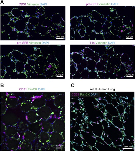
Immunostaining of 3D, multicellular organoids compared with adult human distal lung. (A): Confocal micrograph of cross sections of 3D multicellular lung organoids with immunofluorescence for CD31 (HUVECs), vimentin (FLFs), and pro‐SPB and pro‐SPC (type II alveolar epithelial cells) and T1a (type I alveolar epithelial cells; scale bar = 100 μm). (B): Confocal micrograph of multicellular 3D lung organoids with immunofluorescence for CD31 (HUVECs) and PanCK (SAECs). FLFs were also seeded. (C): Confocal micrograph of a cross‐section of normal adult human lung with immunofluorescence for CD31 (HUVECs) and PanCK (SAECs; scale bar = 100 μm). Abbreviations: 3D, three‐dimensional; DAPI, 4′,6‐diamidino‐2‐phenylindole; FLFs, fetal lung fibroblasts; HUVECs, human umbilical vein endothelial cells; SAECs, small airway epithelial cells; SPB, Surfactant Protein B; SPC, Surfactant Protein C.