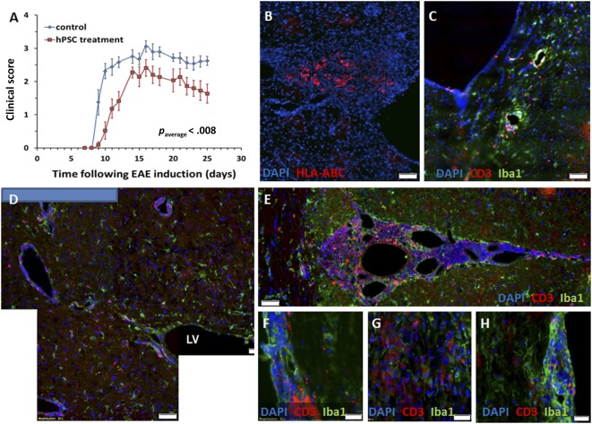
Figure 2.

Intracranial implantation of hPSCs to EAE mice. hPSCs were implanted in the periventricular white matter of mice on day 7 after EAE induction before the development of clinical signs (n = 12 mice). (A): Significant (p = .001) disease attenuation was observed in the clinical course of hPSC‐implanted mice compared with a control EAE group (n = 11). (B): hPSCs were detected 18 days after implantation by HLA‐ABC immunostaining of the mouse brain sections. (C): EAE mice exhibited a typical inflammatory response in their brains on day 25 after EAE induction. (D, E): A strong inflammatory response was noted in the hPSC‐transplanted mice in proximity to the implantation site and in the brain meninges. Both meningeal and parenchymal spinal cord infiltrates were detected in the EAE mice (F, G), but the hPSC‐implanted mice exhibited mostly meningeal infiltrates (H). Scale bars = 50 µm (B–E) and 20 µm (F–H). Abbreviations: DAPI, 4′,6‐diamidino‐2‐phenylindole; EAE, experimental autoimmune encephalomyelitis; HLA, human leukocyte antigen; hPSC, human placental stromal cell; LV, lateral ventricle.
