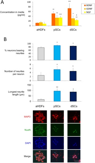
Figure 3.

Directly converted Schwann cells (dSCs) significantly produced neurotrophic factors and induced neurons to produce neurites (A) Cells (2 × 10⁵ per well) were cultured for 48 hours, and the conditioned medium was analyzed by ELISA to determine the concentrations of the indicated neurotrophic factors. The data are expressed as means ± SD (n = 6 experiments). **p < .01, ***p < .001 versus normal human dermal fibroblasts (aHDFs), # p < .05 versus pSCs. (B): The indicated cells were cocultured with NG108‐15 neuronal cells for 24 hours. NG108‐15 cells were immunostained with anti‐MAP2 and anti‐NeuN antibodies, followed by Alexa 488‐ and Alexa Fluor 546‐conjugated secondary antibodies to visualize the neurites. Cell nuclei were stained with DAPI. Representative fluorescent microscopic images are shown (lower) (Scale bar = 50 µm). The percentage of neurite‐bearing neurons, the number of neurites per neurons, and the longest neurite length were calculated using ImageJ software (20 cells were analyzed for each group) (upper). The data are expressed as means ± SD (n = 4 cultures), *p < .05, **p < .01 versus aHDF culture. The experiments were repeated three times. Abbreviations: aHDFs, normal human dermal fibroblasts; BDNF, brain‐derived neurotrophic factor; dSCs, directly converted Schwann cells; GDNF, glial cell‐derived neurotrophic factor; NGF, growth factor.
