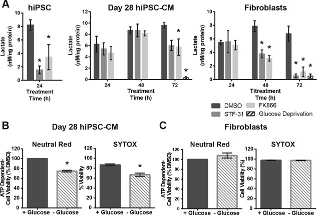
Figure 4.

Nicotinamide phosphoribosyltransferase inhibition affects glycolytic flux in a cell type dependent manner. (A): Lactate secretion as measured by colorimetric assay following treatment with 2.5 µM STF‐31 or 100 nM FK866 for 24 hours in hiPSC and 24–72 hours in day 28 hiPSC‐CM and fibroblasts. (B, C): Bar graphs depicting cell viability following 72 hours of glucose deprivation as measured by neutral red and SYTOX cell death assay in day 28 hiPSC‐CM (B) and fibroblasts (C). Data are represented as mean ± SEM for 3–6 biological replicates in each group (N = 3 STF‐31 and FK866; N = 6 DMSO for hiPSC and fibroblasts, N = 3 cardiomyocytes). *, p < .05. Abbreviations: DMSO, dimethyl sulfoxide; hiPSC‐CM, human induced pluripotent stem cells‐derived cardiomyocytes.
