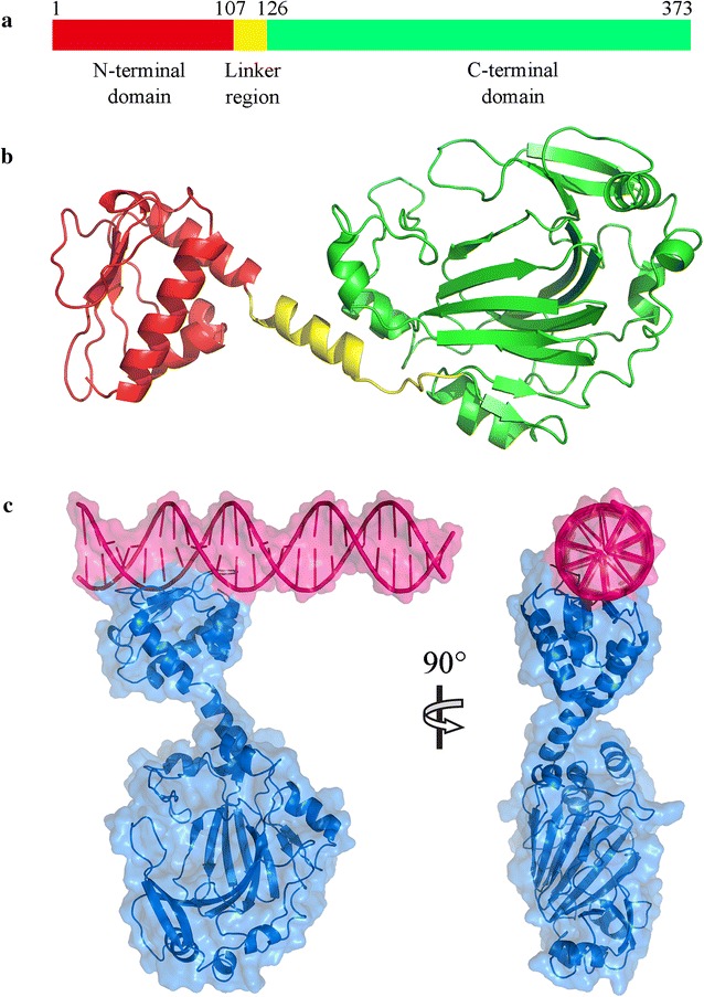
Fig. 4.

The structure modeling of Esi protein and the DNA-Esi binding complex. a Positions of N-terminal domain, linker region and C-terminal domain. b The structure model of Esi protein. The protein backbone is shown in Cartoon mode. There were three parallel β-pleated sheets and three alpha helixes in the N-terminal domain and an alpha helix in the linker region. The C-terminal region is a cupin-like domain. c The DNA–protein complex structure of Esi protein (blue) and epothilone promoter (red). The top of the N-terminal domain of Esi binds with the promoter DNA sequence
