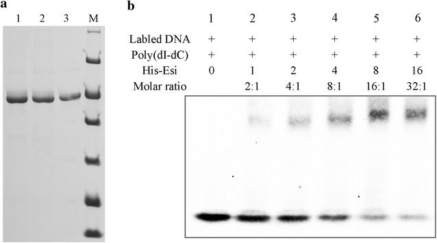
Fig. 5.

a The purified Esi protein with His-tag was detected by SDS-PAGE and visualized with Coomasie brilliant blue staining. Lanes 1, 2, and 3 represented purified His-Esi protein; Lane M represented molecular mass standards (from top to bottom, 116, 66.2, 45, 35, 25, 18.4, 14.4 kDa). b EMSA showing the binding of His-Esi protein to the 17-bp operator DNA sequence of epothilone promoter. Lane 1 represented free DNA with no His-Esi protein; Lanes 2 to 6 represented DNA incubated with increased concentrations of His-Esi
