
Fig. 3.
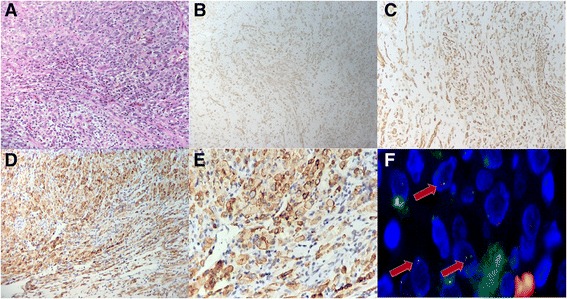
a. Tumors were composed of both large epithelioid cells and spindle cells. b, c, d. The tumor cells were positive for SMA (B), desmin (C) and ALK (D). e. IHC for ALK showed positive staining in both large epithelioid cells and spindle cells. The staining was most distinctive under the membranes. f. Fluorescence in situ hybridization showing splitting of the two signals corresponding to the 3′ and 5′ ends of ALK, confirming the presence of an ALK rearrangement