Abstract
Injuries to the midtarsal joints usually occur in various combinations such as fracture, fracture subluxation, and fracture dislocation. Isolated dislocations of the navicular bone without fracture are rare injuries. The few existing case reports describe the probable mechanism of injury and optimal treatment. We present a 64-year-old diabetic man whose tarsal navicular was completely dislocated without fracture following a traffic accident. The most probable mechanism of injury was an abduction–pronation injury causing a midtarsal dislocation, and on spontaneous reduction, the navicular was dislocated medially. This mechanism is similar to perilunate dislocation. Computed tomography of the involved foot was done to accurately define the full extent of the bony injury and magnetic resonance imaging was required to determine if there was a ligamentous injury and to assess the attachment of soft tissues to the displaced bone to help assess the risk of avascular necrosis. The patient was treated successfully with open reduction and primary talonavicular arthrodesis with Kirschner wires.
Keywords: Midtarsal injuries, Navicular dislocation, Talonavicular arthrodesis
1. Introduction
The tarsal navicular bone is strategically located in the uppermost portion of the medial longitudinal arch of the foot, and hence, plays a major role in weight bearing during ambulation. It acts as the keystone for vertical stress on the arch. Multiple deforming forces act on the navicular during various sporting activities and dancing and in high energy injuries resulting in varied degrees of fracture, subluxation or dislocation involving the tarsus and metatarsus. Pure navicular dislocation is a rare injury. We could find only ~15 reported cases [1,2,3,4,5,6]. Because of its rarity, the mechanism of injury is poorly understood and optimum treatment is difficult to plan. All reported cases were associated with other, bony or ligamentous injuries [7]. A few cases were treated with closed reduction while open reduction and Kirschner wire fixation were used in others. Our reported patient had peripheral diabetic neuropathy and features of arthropathy, and hence treatment consisted of open reduction and primary arthrodesis of the talo-navicular joint.
2. Case report
A 64-year-old man presented with injury to the right foot from a traffic accident. He had swelling over the dorsomedial aspect of the foot (Fig. 1). There were no external wounds. The patient had significantly less pain than most patients with this type of the injury. He had a history of diabetes and had taken insulin and oral hypoglycemics for >10 years. Clinical examination revealed swelling of the bone and diabetic peripheral neuropathy.
Fig. 1.
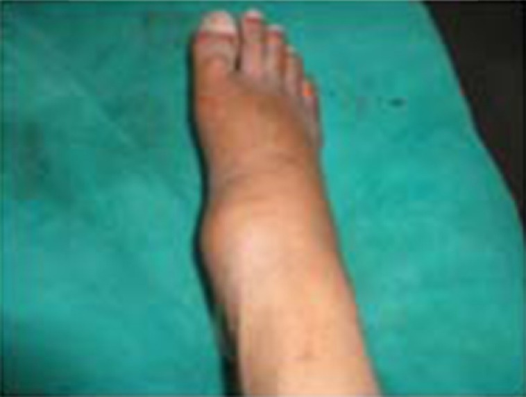
Clinical appearance of the injury.
2.1. Investigation
Radiographs showed complete dislocation of the navicular without fracture. A lateral radiograph revealed a gap between the talus and the medial cuneiform (Fig. 2). The navicular was superomedial to the head of the talus. Computed tomography of the foot showed no fracture of the navicular or other tarsal bones and confirmed the diagnosis (Fig. 3). Blood investigations revealed that his glucose levels were within normal limits. Magnetic resonance imaging of the foot was desirable to determine if there was injury to ligamentous structures, which allows the navicular to dislocate without fracture.
Fig. 2.
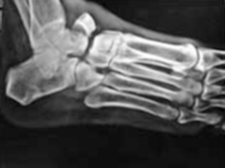
Preoperative radiograph showing a displaced navicular.
Fig. 3.
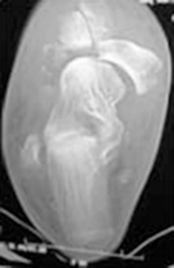
Preoperative computed tomography showing dislocated navicular.
2.2. Management
The treatment plan included talonavicular arthrodesis because the patient had associated diabetic arthropathy. The joint was approached through a dorsomedial approach. The navicular was displaced completely and was devoid of any soft tissue attachment (Fig. 4). It was denuded of its articular cartilage, reduced in its anatomical position, and fixed in place with two Kirschner wires. The wound was closed in layers after hemostasis, and the ankle was immobilized in a below-knee posterior slab.
Fig. 4.
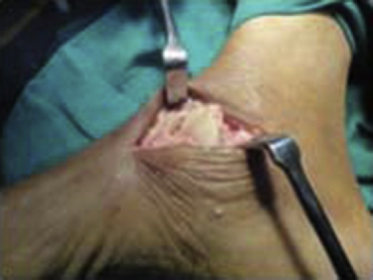
Peroperative photograph of the displaced navicular.
2.3. Follow-up
The sutures were removed at 2 weeks and immobilization of the ankle and foot was continued for 3 months. Gradual weight bearing was then started. Postoperative radiographs showed a well-reduced navicular held in position with two Kirschner wires (Fig. 5). The patient showed excellent clinical results. The patient was lost to follow-up shortly thereafter.
Fig. 5.
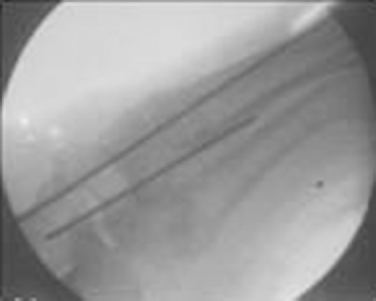
Postoperative radiograph showing the navicular held in place with Kirschner wires.
3. Discussion
3.1. Functional anatomy
The tarsal navicular is a boat-shaped bone. It articulates distally via three facets with the medial, intermediate and lateral cuneiforms. Because of its proximal articulation with the talar head, the talonavicular joint has the greatest range of motion, and along with the calcaneocuboid joint forms Chopart's joint.
The bones of the midfoot fit snugly against one another and are shaped to form the transverse and longitudinal arches. The medial longitudinal column consists of the talus, the navicular, the three cuneiforms and the corresponding three metatarsals. The lateral column consists of the calcaneus, the cuboid and the corresponding two metatarsals. The navicular is the keystone of the medial longitudinal arch, and is rigidly stabilized by an extensive network of dorsal and plantar ligaments [8]. The rarity of this injury can be attributed to this surrounding rigid bony and ligamentous support. The navicular usually undergoes fracture and dislocation rather than pure dislocation [1].
The blood supply to the navicular comes from small branches of the posterior tibial and dorsalis pedis arteries. The proximal and distal portions of the navicular are covered by articular cartilage and the blood supply enters both medially and laterally. The central third portion is therefore relatively avascular. When devoid of surrounding soft tissues, as in the case of complete dislocation, it is prone to avascular necrosis.
3.2. Mechanism of injury
The navicular plays a major role in weight bearing during ambulation as a result of its strategic location in the medial longitudinal arch of the foot. It acts as the keystone for vertical stress on the arch. Because of the complexity of the midtarsal and tarso-metatarsal joint complex, the exact mechanism of injury [2,9] is often not known, particularly when there are multiple deforming forces present, as in high-energy injuries. The following sports and activities have a relatively high risk of navicular injury: (1) sports involving jumping and sprinting – basketball, soccer, football, and rugby; (2) ballet and other dancing activities, and gymnastics; (3) biomechanical abnormality; and (4) military training.
3.3. Classification
Broadly two categories of injury, high energy and low energy, occur in the midtarsus involving the navicular. High-energy injuries are a result of motor vehicle collisions, falls from a height, and crush injuries. Low-energy injuries are those due to twisting injuries in athletes.
Broca and Malgaigne (1852–56) classified talocalcaneal navicular joint dislocations, according to the direction of dislocation and their frequency with medial dislocations most frequently encountered, followed by lateral, posterior, and anterior dislocations. Medial talocalcaneal navicular dislocations are often called “acquired clubfoot” (because of their appearance) or “basketball foot” (a term coined by Graham in 1964), because many of these injuries are associated with sport, particularly basketball. Lateral talo-calcaneal navicular dislocations are often called “acquired flatfoot”.
3.4. Clinical examination
The clinical symptoms and signs of midfoot injuries can vary and be mild, especially following spontaneous reduction of dislocations, preventing early diagnosis: (1) swelling over the dorsomedial aspect of the foot; (2) tenderness at the “N spot”, which is defined as the proximal dorsal portion of the navicular; and (3) pain with active inversion and passive eversion. Examination under anaesthesia and stressing the midfoot with an abduction and pronation stress test may reveal instability [10].
3.5. Investigations
For all midfoot injuries, standard anteroposterior, lateral and oblique radiographs should be obtained. (1) Radiographs: the continuity of the cortical bone should be examined for a fracture. Its displacement and its relation with the surrounding bones are noted to determine subluxation or dislocation. (2) Technetium 99m bone scans: increased radionuclide uptake indicates a navicular stress fracture. (3) Computed tomography: confirms the diagnosis of navicular fractures and relative positions of other tarsal bones. (4) Magnetic resonance imaging: modality of choice for diagnosing ligamentous injury [11] and vascular insult to the navicular.
3.6. Management
The main aim of treatment is early stable anatomical reduction. Anatomical reduction whether achieved via open or closed means is probably dependent on several factors, including timing of surgery, presence of fracture fragments, and soft tissue interposition (tibialis anterior tendon) preventing anatomical reduction. Preservation of the longitudinal arch is necessary for a good clinical outcome. Early reduction reduces the risk of vascular compromise.
Dislocations that are complicated by fractures are assessed for stability following reduction. If the navicular is stable, then treatment includes a non-weight-bearing cast for 6 weeks. If the navicular is unstable, then internal fixation is required. After 6 weeks of non-weight bearing, the patient is assessed for pain at the N spot. If the patient is free of pain, a gradual return to normal activity is begun. This gradual return should be in a stepwise fashion over 6 weeks. In cases of persistent pain at the navicular, a custom-molded orthotic with longitudinal and transverse arch support may be prescribed to help relieve stress on the navicular.
The fixation method varies from using screws, plates, Kirschner wires or external fixators. There are concerns that Kirschner wires can loosen and become infected during a long duration of fixation. The potential advantages of plate fixation rather than transarticular screw fixation are less damage to the joint surface and a theoretically decreased incidence of post-traumatic arthritis. After fixation, the postoperative protocol remains the same as for conservative management.
The functional outcome regarding pain in long-term follow-up appears to be better with primary arthrodesis. Primary arthrodesis reduces the need for repeat surgery compared with internal fixation.
3.7. Complications
These can be early or delayed and depend on several factors including the type of dislocation, severity of the injury, presence of associated fractures, and postoperative course. Complications are: (1) prolonged disability due to persistent pain in the navicular; (2) stiffness of the midfoot; (3) nonunion of associated fractures; (4) avascular necrosis of the navicular; (5) deformity of the foot; and (6) post-traumatic arthritis. Some of these complications may require a second surgical procedure in the form of arthrodesis or excision of the navicular.
Footnotes
Conflict of interest: none.
References
- [1].Vaishya R, Patrick JH. Isolated dorsal fracture dislocation of the tarsal navicular. Injury. 1991;22:47–8. doi: 10.1016/0020-1383(91)90162-8. [DOI] [PubMed] [Google Scholar]
- [2].Pathria MN, Rosenstein A, Bjorkengren AG, Gershuni D, Resnick D. Isolated dislocation of the tarsal navicular: a case report. Foot Ankle. 1988;9:146–9. doi: 10.1177/107110078800900311. [DOI] [PubMed] [Google Scholar]
- [3].Freund KG. Isolated dislocation of the tarsal navicular. Injury. 1989;20:117–8. doi: 10.1016/0020-1383(89)90157-5. [DOI] [PubMed] [Google Scholar]
- [4].Hooper G, Hughes S. Midfoot and navicular injuries. In: Helal B, Wilson D, editors. The foot. Edinburgh: Churchill Livingstone; 1988. pp. 932–43. [Google Scholar]
- [5].Davis AT, Dann A, Kuldjanov D. Complete medial dislocation of the tarsal navicular without fracture: report of a rare injury. J Foot Ankle Surg. 2013;52:393–6. doi: 10.1053/j.jfas.2013.01.001. [DOI] [PubMed] [Google Scholar]
- [6].Mathesul AA, Sonawane DV, Chouhan VK. Isolated tarsal navicular fracture dislocation: a case report. Foot Ankle Spec. 2012;5:185–7. doi: 10.1177/1938640012439602. [DOI] [PubMed] [Google Scholar]
- [7].Dhillon MS, Nagi ON. Total dislocations of the navicular: are they ever isolated injuries? J Bone Joint Surg Br. 1999;81:881–5. doi: 10.1302/0301-620x.81b5.9873. [DOI] [PubMed] [Google Scholar]
- [8].Early JS, Hansen ST., Jr . Midfoot and navicular injuries. In: Helal B, Rowley DI, Cracchiolo A III, Myerson M, editors. Surgery of disorders of the foot and ankle. London: Martin Dunitz; 1996. pp. 731–47. [Google Scholar]
- [9].Dixon JH. Isolated dislocation of the tarsal navicular (letter) Injury. 1979;10:251. doi: 10.1016/0020-1383(79)90022-6. [DOI] [PubMed] [Google Scholar]
- [10].Myerson MS. The diagnosis and treatment of injury to the tarsometatarsal joint complex. J Bone Joint Surg. 1999;81:756–63. doi: 10.1302/0301-620x.81b5.10369. [DOI] [PubMed] [Google Scholar]
- [11].Preidler KW, Peicha G, Laitai G, Seibert FJ, Fock C, Szolar DM, et al. Conventional radiography, CT, and MR imaging in patients with hyperflexion injuries of the foot: diagnostic accuracy in the detection of bony and ligamentous changes. AJR Am J Roentgenol. 1999;173:1673–7. doi: 10.2214/ajr.173.6.10584818. [DOI] [PubMed] [Google Scholar]


