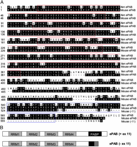
Fig. 1.
Amino acid sequences and domain structures of mouse ePAB alternatively spliced forms. (A) Pairwise alignment of the mouse and the Xenopus ePAB amino acid sequences. The sequences of both ePAB spliced variants, including and excluding exon 11, are shown. The four RRM motifs are boxed in red, and the PABP domain is boxed in blue. (B) Schematic representation of mouse ePAB alternatively spliced forms. The four RRMs are indicated by gray boxes. Because the PABP domain is encoded by exons 10.7 and 11, the shorter form (-ex 11) contains only part of the PABP motif (amino acids 524–543). The C termini of the two forms also differ because of shifting of the exon 12 reading frame.

