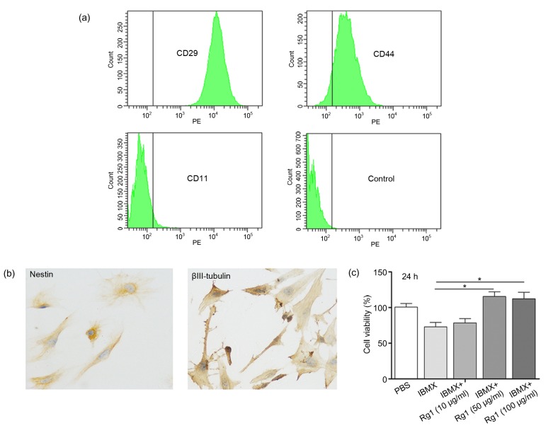
Fig. 1.
Effect of ginsenoside Rg1 on cell proliferation during ADSC neural differentiation
(a) Isolated ADSC were analyzed by flow cytometry. Cells were positive for ADSC markers CD44 and CD29, and negative for CD11b, a hematopoietic cell surface marker. PE: phycoerythrin. (b) Immunohistochemical staining of ADSC after neural induction. (c) ADSC were treated with different concentrations of ginsenoside Rg1 during IBMX induction, and cell viability was determined by MTT assay. Values are expressed as mean±standard deviation (SD) of triplicate experiments. * P<0.05

