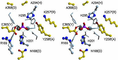
Fig. 2.
Stereodiagram of the active-site residues in sweet potato PAP. Side chains that coordinate directly to the metal ions are shown as stick models. Side chains that form the active site are shown as ball-and-stick models. Carbon atoms colored gray represent side chains that are conserved between red kidney bean and sweet potato PAP. Carbon atoms colored yellow are nonconserved, and the name of the equivalent residue in red kidney bean PAP is in brackets. Primed amino acid residues are from the neighboring subunit.

