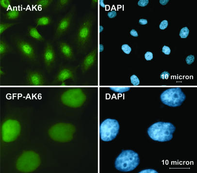
Fig. 3.
Subcellular localization of AK6 in HeLa cells by means of fluorescence microscopy. (Upper Left) AK6 was detected in vivo by rabbit anti-AK6 and goat anti rabbit IgG-FITC. (Upper Right) DAPI staining on the same cells confirmed a nuclear localization of AK6. (Lower Left) HeLa cells were transfected by pEGFP-AK6. (Lower Right) After transfection (24 h), cells were visualized with a fluorescent microscope, and strong nuclear fluorescence was observed. (Lower Right) DAPI staining on the same cells again proved the nuclear localization of AK6.

