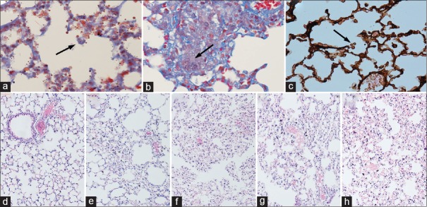
Figure 1.
Histology of lung tissue stained with H and E, Masson trichrome, and PASM. (a–c) The fungal spores of Aspergillus fumigatus. (d) Control animal. (e) Animal infected with IPA. (f) Immunosuppression plus aspergillosis group (CTX + IPA). (g) Immunosuppression plus IPA plus IL-12 treatment group (CTX + IPA + IL-12). (h) Immunosuppression plus aspergillosis plus rapamycin treatment group (CTX + IPA + RAPA). Original magnification: (a) Masson trichrome staining, original magnification ×200; (b) Masson trichrome staining, original magnification ×400; (c) PASM staining, original magnification ×600; (d–h) H and E, original magnification ×100. CTX: Cyclophosphamide; IPA: Invasive pulmonary aspergillosis; PASM: Periodic acid silver methenamine; IL: Interleukin.

