
Figure 3.
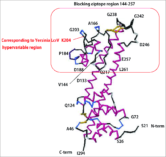
Predicted tertiary structure of PcrV. Based on the structural information for LcrV (Swissplot IR6F), the tertiary structure of PAO1 PcrV was predicted by the protein structure prediction server RaptorX.58 PcrV, consisting of 294 amino acids, forms a central shaft structure, with coiled-coil double strands and 2 globular domains at either end. The C-terminal globular domain includes amino acid 204, corresponding to the hypervariable region of Yersinia LcrV, and amino acids 144–257, recognized as the mAb166 blocking epitope.