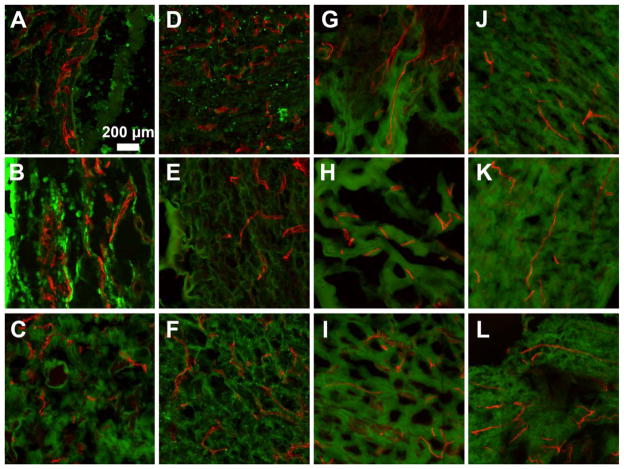
Figure 9.
Representative lectin stained images of hydrogel implants harvested at week 1 (A–F) and week 3 (G–L). (A–C, G–I) Representative images of hydrogels modified with LGPAS, M2S, M14S peptide substrates at weeks 1 and 3, respectively. (D–F, J–L) Representative images of hydrogels modified with LGPAT, M2T, and M14T peptide substrates at weeks 1 and 3, respectively.

