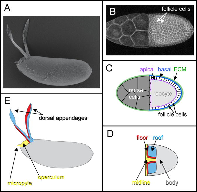
Figure 1.

The formation of Drosophila eggshells from egg chambers is a model for epithelial patterning and morphogenesis. A) Scanning Electron Micrograph (SEM) of an eggshell from Drosophila melanogaster. B) A stage-10 D. melanogaster egg chamber, visualized with a fluorescent membrane marker. In the right half of the image, the columnar follicle cells surround and thus obscure the underlying oocyte. In the left half of the egg chamber are 15 nurse cells; these are covered by a thin layer of squamous “stretch” follicle cells that are not readily apparent under these visualization conditions. C) Schematic of a stage-10 egg chamber, in cross-section. The stretch cells, not shown, do not produce eggshell material. D) Schematic of a stage-10 egg chamber (dorsal view), showing the four distinct cell types of columnar follicle cells. E) Schematic of an eggshell (lateral view), color-coded to show which eggshell structures are formed by which follicle-cell type in (D). Figures C-E are adapted from (Osterfield et al., 2015).
