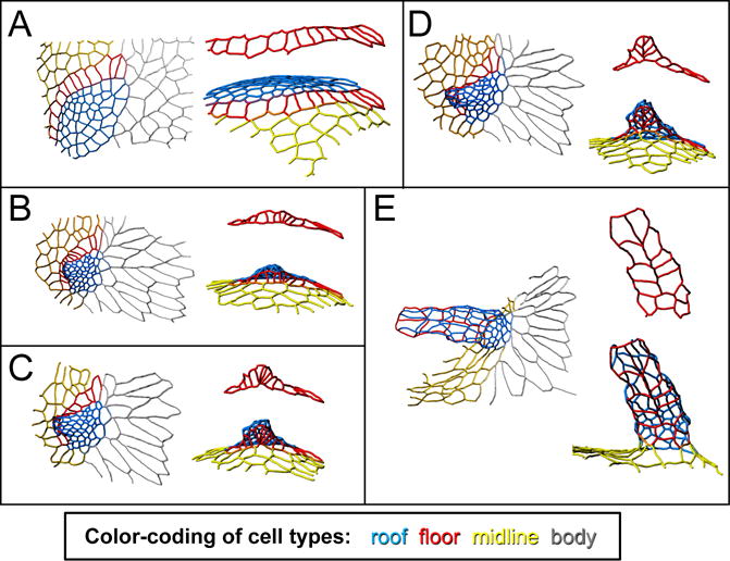
Figure 3.

Changes in cell shape and neighbor relationships transform a flat appendage primordium into a dorsal-appendage tube. The tracings in these panels show the apical outlines of a patch of follicle cells, in their transformation from a flat primodium (A) to a fully formed, though not fully elongated, dorsal-appendage tube (E). Color-coding as in Figure 1: blue is roof, red is floor, yellow is midline (operculum), and gray is body. For each time point, the left image shows a dorsal view, while the right image is a view from below the appendage, i.e. a roughly ventral-anterior view. Of particular interest are the floor cells (also shown by themselves in red), which “zip” to form the two-cell wide appendage floor. This figure is adapted from (Osterfield et al., 2015).
