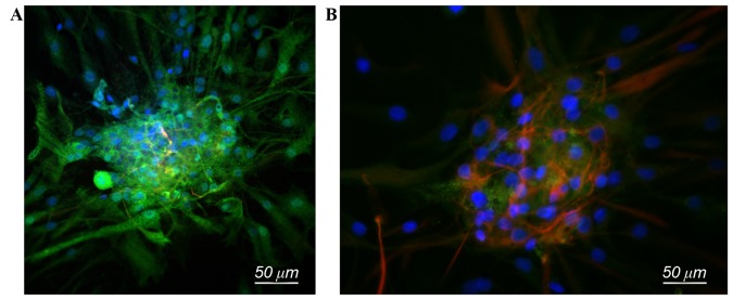
Figure 2.
Immunofluorescence staining of co-cultured and NSC groups. (A) NSC neurospheres in the co-cultured group gradually differentiate into neurons with Map2-positive immunofluorescence staining (green), cell nuclei stained by DAPI (blue). (B) NSC group, a small number of cells differentiate into neurons, Map2-positive (green), while a large proportion of cells differentiate into astrocytes, glial fibrillary acidic protein positive (red), cell nuclei stained by DAPI (blue). Scale bar, 50 µm. NSC, neural stem cells; Map2, microtubule associated protein; DAPI, 4′,6-diamidino-2-phenylindole.

